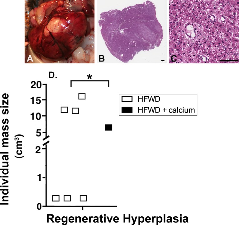
Fig 3. Regenerative hyperplasia (RH).
Representative RH lesions are shown grossly (A) and histologically (B and C). RH nodules were distinguished from neoplastic masses by preservation of hepatic structures (ie. portal triads, central veins, 1–2 cell thickness of hepatic cords) within the area of proliferation. D. Frequency of RH nodules. Six of 20 HFWD mice had liver masses histologically classified as RH compared to 1 of 20 HFWD-Ca mice (Chi-square, 2-tailed, p = 0.0375). (Scale bars = 1mm [B] and 100μm [C]).

