Figure 5.
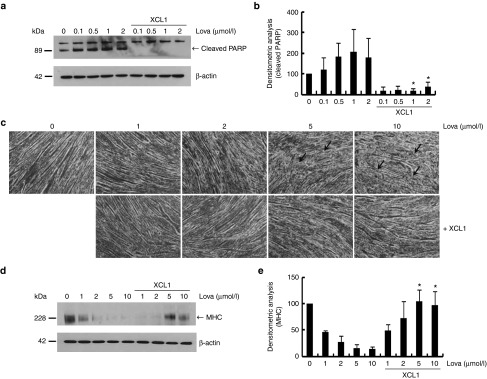
XCL1 treatment inhibited lovastatin-induced apoptosis of C2C12 cells and reduction of myotube formation. (a) C2C12 cells were treated with lovastatin (0.1, 0.5, 1, and 2 μmol/l) in a dose-dependent manner in the absence or presence of XCL1 and protein lysates were analyzed by Western blot analysis using anti-poly ADP-ribose polymerase (PARP) antibody. (b) Cleaved PARP fragments were monitored by densitometric analysis (*P < 0.05, n = 3). (c) The 7-day differentiated myotubes from C2C12 cells were treated with lovastatin (1, 2.5, 5, and 10 μmol/l) in the absence or presence of XCL1. Each arrow indicates defects in the myotubes. (d,e) Protein extracts of myotubes were analyzed by Western blot analysis using anti-myosin heavy chain (MHC) antibody. Bands of MHC were monitored by densitometric analysis (*P < 0.05, n = 3).

