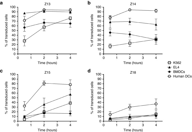
Figure 3.
Comparison of the transduction capacity of Z13, Z14, Z15, and Z18 CPP truncations. Transduction was assessed in cells with high phagocytosis capacity (DCs of human and mice origin) and in cells with poor phagocytosis capacity (human K562, or murine EL4). Cells were incubated for 30 minutes, 2 hours or 4 hours with the fluorescein-conjugated constructs (Z13OVACD8FAM, Z14OVACD8FAM, Z15OVACD8FAM or Z18OVACD8FAM) then subjected to a 30 seconds wash with an acidic buffer to remove membrane bound peptide before staining for flow cytometry analysis. Mean and SEM of three independent experiments are shown. DC, dendritic cells; CPP, cell penetrating peptide; OVA, ovalbumin.

