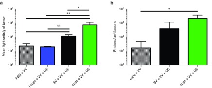
Figure 1.
In vivo infectivity of vaccinia virus (VV) delivered using SonoVue (SV) or polymeric cup (“cups”) nucleated cavitation. A dose of 1 × 10⁵ luciferase expressing VV was mixed with inactive cups (i-cups), SV or cups and injected into (a) Balb/c mice bearing CT-26 tumors. The tumors were exposed to ultrasound (US) (see Methods for parameters) and 48 hours later tumors were excised, homogenized and luciferase expression quantified (see Methods). (b) CD-1 nude mice bearing HepG2 tumors. The tumors were exposed to US and 5 days later luciferase expression was assessed by an in vivo imaging system (IVIS) (see Methods). n = 4, SD shown, significant differences (*P < 0.05, **P < 0.01, ns, nonsignificant P > 0.05) detected by one-way analysis of variance (ANOVA) with Tukey compare all columns post-test.

