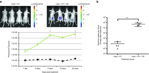
Figure 2.
In vivo infectivity of vaccinia virus (VV) delivered using polymeric cup (“cups”) nucleated cavitation to HepG2 tumours. A dose of 1 × 10⁵ luciferase expressing VV was mixed with cups and injected into mice and their tumors exposed to ultrasound (US) (see methods for parameters). Passive acoustic mapping confirmed the absence or presence of cavitation within the tumor. (a) Luciferase expression was assessed by an in vivo imaging system (IVIS) imaging at intervals over the next 20 days (see Methods, inset images show luciferase expression of tumours at day 10). Green line = cups + VV + US, black dashed line = cups + VV. (b) VV genome copy number within the tumors of the mice was measured at sacrifice on day 20 (see methods). n = 4, SD shown, significant differences (*P < 0.05, **P < 0.01) detected by analysis of variance (ANOVA) with Bonferroni compare all columns post-test.

