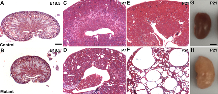
Figure 1. Tissue sections and gross images show progressive cyst formation in Six2creFrs2αKO kidneys.
(A–F,H,E) Stained sections of control (A,C,E) and Six2creFRS2αKO (B,D,F) kidneys show that the mutants are hypoplastic at E18.5 (A vs. B), develop small cysts dispersed in normal appearing parenchyma at P7 (C vs. D), and have cysts replacing most of the parenchyma by P21 (E vs. F). (G,H). Dissected kidneys from P21 control (G) and Six2creFRS2αKO (H) mice show that the mutant kidneys are enlarged and have multiple fluid-filled cysts. (A–F) scale bar = 200 μm; G, H scale bar = 0.25 cm.

