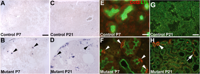
Figure 6. Ccl2 is ectopically expressed initially in non-dilated Six2creFrs2αKO proximal tubular cells.
(A,B) At P7, in situ hybridization reveals no apparent Ccl2 expression in controls (A), but diffuse punctate staining exclusively in non-dilated cortical tubular cells in mutants (B, arrowheads). (C,D) At P21, in situ hybridization again shows no Ccl2 staining in controls (C), but reveals a larger number of mostly non-dilated mutant cortical cells with ectopic expression of Ccl2 (D, arrowheads). (E,F) At P7, co-immunofluorescence for LTL (green) and Ccl2 (red) reveals no Ccl2 expression in controls (E), but ectopic Ccl2 expression in individual cells within non-dilated LTL-positive proximal tubules in mutants (F, arrowheads). (G,H) At P21, co-labeling immunofluorescence reveals no signal in controls (G), but strong labeling of minimally-dilated LTL-positive tubules in the outer cortex (arrowhead) and of individual cells within LTL-positive cyst lining cells (arrow). (A–D) scale bar = 100 μm; (E,F) scale bar = 12.5 μm; (G,H) scale bar = 25 μm.

