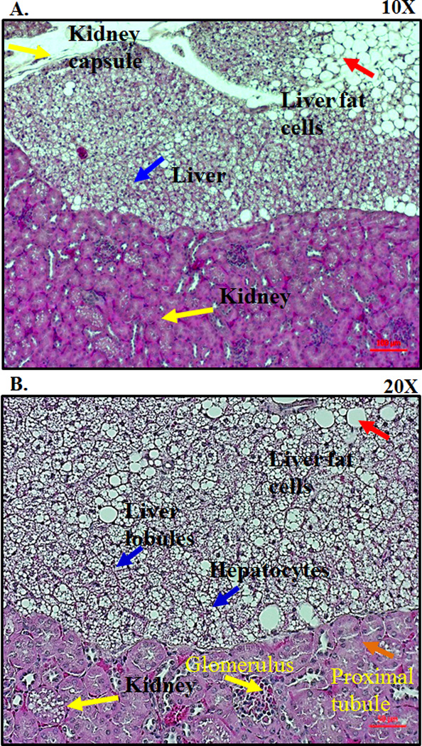
Figure 1.

Histological analysis of the transplanted liver tissue under the kidney capsule. MASP-1/3−/−(recipient) mouse was transplanted with the fD−/− (donor) mouse liver tissue under the kidney capsule. All mice were sacrificed at 5–14 weeks (n = 5). Kidneys, both with (left organ) and without (right organ) transplanted liver fragments, were removed and fixed in a 10% neutral buffered formalin. Histology sections from the kidneys were obtained from all mice to assess for viable liver tissue. A. Hematoxylin and eosin staining from one transplanted mouse at 5-weeks showing the presence of healthy hepatocytes (blue arrow) under the kidney capsule (yellow arrow) at a 10X magnification. Presence of adipose tissue or fat cells (red arrow) in the liver tissue as well as under the kidney capsule at a 10X magnification. B. Presence of hexagonal type hepatocytes (blue arrow) with dividing nucleus under the kidney capsule at a 20X magnification. Presence of kidney capsule showing glomerulus (yellow arrow) and proximal renal tubule (orange arrow) at a 20X magnification. Red scale bars in A and B equals 0.1 mm (100 µm).
