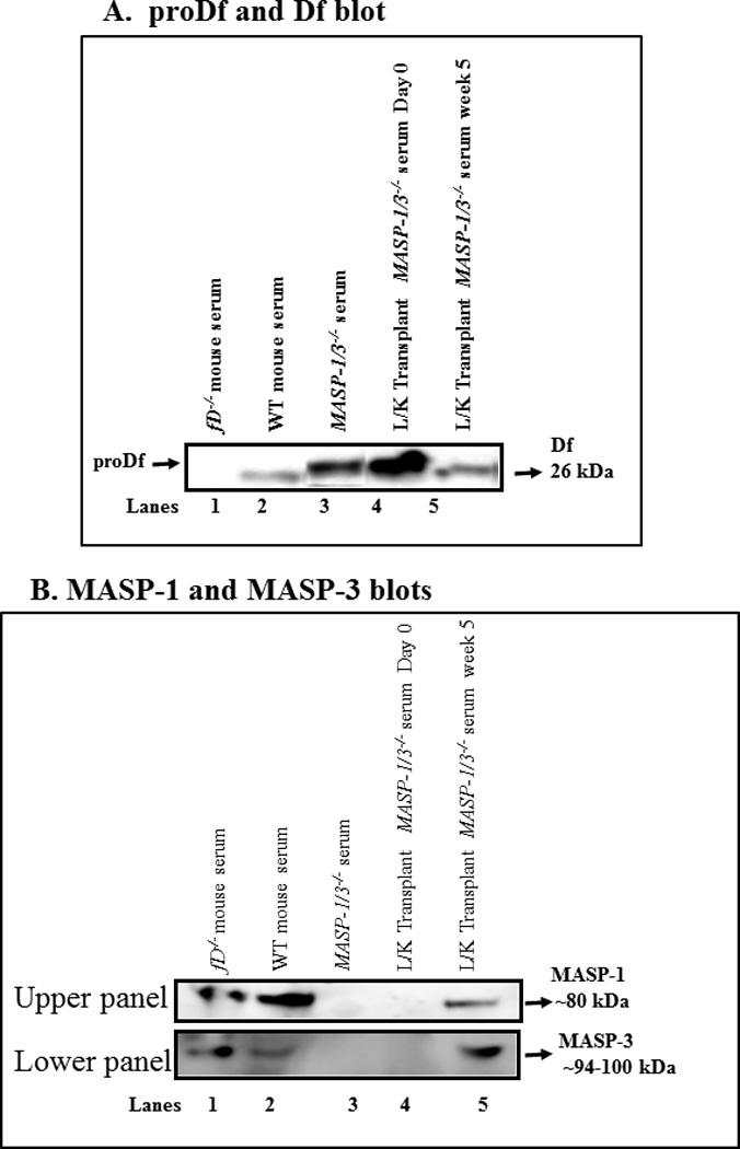
Figure 2.

Western blot analysis of the cleavage of the proDf into Df and hepatocyte-specific MASP-1 and MASP-3 proteins generated in the circulation. MASP-1/3 cleavage of proDf results in a loss of 5 amino acids which can be visualized as a slight increase in mobility in SDS-PAGE. Sera from MASP-1/3−/− (receipt) mice were obtained before day 0 and 5 weeks following liver transplantation. Serum from fD−/− (donor) mouse mice were also obtained. A. To examine the cleavage of proDf into Df protein in the circulation of MASP-1/3−/− mice, before and after liver transplantation, sera were immunoprecipatated for Df and then digested with PNGase F overnight at 37°C, according to Material and Methods. A 12% Bis-Tris reducing SDS-PAGE gel was used to distinguish between proDf and Df proteins. Sera from fD−/− and WT mice were used as negative and positive controls (lanes 1, 2). A band of proDf was still intact in the serum from a MASP-1/3−/− mouse (lane 3) and also in MASP-1/3−/−(recipient) mice prior to liver transplantation (lane 4). A clear Df band of 26 kDa was present in the serum of a MASP-1/3−/− mouse 5 weeks after liver transplantation (lane 5), indicating the cleavage of proDf into Df. The presence of MASP-1 and MASP-3 proteins in the circulation of MASP-1/3−/− mice transplanted with the liver under the kidney capsule was confirmed by using mannose and N-acetyl-D-glucosamine, respectively, pull-down assays followed by Western blot analysis as described in the Methods section. B (upper panel) Absence of MASP-1 before liver transplantation in the serum from a MASP-1/3−/− mouse as expected (lane 4) but its presence 5 weeks after liver transplantation in a MASP-1/3−/− mouse (lane 5). Sera from fD−/− and WT mice were used as positive controls (lanes 1, 2), and serum from a MASP-1/3−/− mouse was used as a negative control (lane 3). B (lower panel) Absence of MASP-3 before liver transplantation in the serum from a MASP-1/3−/− mouse as expected (lane 4) but its presence 5 weeks after the liver transplantation in a MASP-1/3−/− mouse (lane 5). Sera from fD−/− and WT mice were used as positive controls (lanes 1, 2), and serum from a MASP-1/3−/− mouse was used as a negative control (lane 3).
