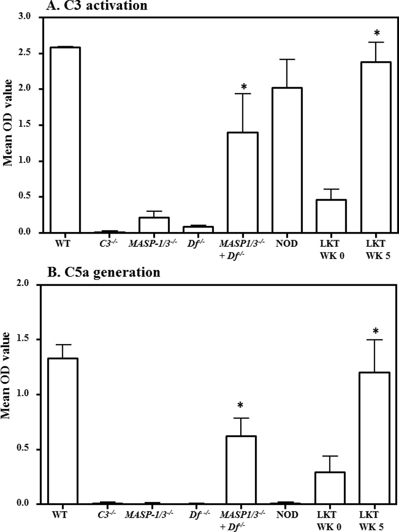
Figure 3.

ELISA demonstrating the restoration of defective AP activity in liver transplanted MASP-1/3−/− mice. The AP activity was examined by ELISA after incubation of serum on adherent anti-collagen antibodies followed by measurement of C3 deposition and C5a generation in a Ca++ deficient buffer (Mg++/EGTA). A. C3 deposition using sera from transplanted MASP-1/3−/− mice before (WK 0) and after transplantation (WK 5). Sera from WT and C3−/− mice were used a positive and negative controls, respectively. Sera from non-transplanted MASP-1/3−/− and fD−/− mice were also used to show the defective AP followed by a full restoration of the defective AP in MASP-1/3−/− mice when sera were mixed (1:1 ratio). B. C5a generated after incubation of sera from transplanted MASP-1/3−/− mice before (WK 0) and after transplantation (WK 5). Sera from WT and NOD mice were used as positive and negative controls, respectively. Sera mixed in vitro from MASP-1/3−/− and fD−/− (1:1 ratio) mice also generated C5a. All data represent the mean + SEM OD values based on n = 4 WT mice; n = 4 C3−/− mice; n = 3 MASP-1/3−/− mice; n = 3 fD−/− mice; n = 3 NOD mice, n = 4 WK 0 transplanted MASP-1/3−/− mice and n = 4 WK 5 transplanted MASP-1/3−/− mice. *p < 0.05 in comparison to the MASP-1/3−/− mice or WK 0. WK = Week
