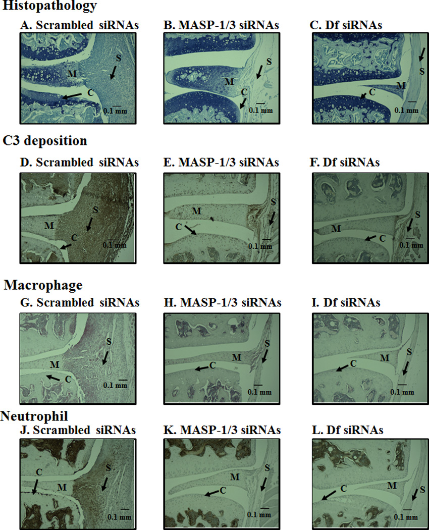
Figure 6.

Representative histopathology, C3 deposition, macrophage and neutrophil images from the knee joints of CAIA mice injected i.p. with scrambled siRNAs or for MASP-1/3 or Df. All joints were fixed with 10% neutral buffered formalin, paraffin embedded, and sectioned at a thickness of 5 µm. The top three panels from left to right (A, B & C) show staining with toluidine-blue (blue color) from the knee joints of CAIA mice treated with scrambled siRNA (left panel) or MASP-1/3 siRNAs (center panel) or Df siRNAs (right panel). The second set of three panels from left to right (D, E & F) show staining with anti-C3 Ab (dark brown color) from the ankle joints of CAIA mice treated with scramble siRNAs (left panel) or MASP-1/3 siRNAs (center panel) or Df siRNAs (right panel). The third set of three panels from left to right (G, H, & I) show staining with F4/80 for macrophages (red color) from the knee joints of CAIA mice treated with scrambled siRNA (left panel) or MASP-1/3 siRNAs (center panel) or Df siRNAs (right panel). The fourth set of three panels from left to right (J, K & L) show staining for neutrophils (brown color) from the knee joints of CAIA mice treated with scramble siRNA (left panel) or MASP-1/3 siRNAs (center panel) or Df siRNAs (right panel). Areas of synovium (S-black arrow), cartilage (C-black arrow), bone (B-black arrow) and meniscus (M-black arrow) are identified. The sections were counterstained with hematoxylin & eosin and photographed under the 10x objective using Zeiss Observer. D1 (AXIO) microscope. Red scale bar in A-L equals 0.1 mm (100 µm).
