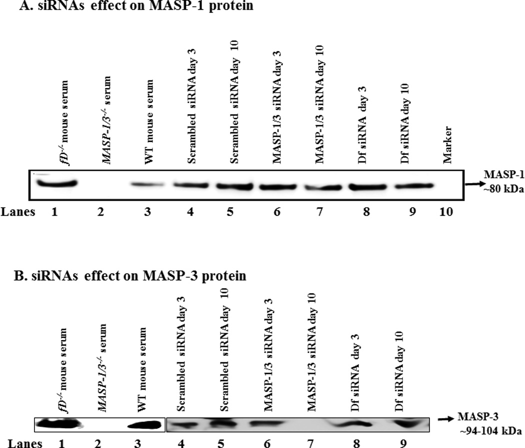
Figure 7.

In vivo efficiency of the siRNAs for MASP-1/3 was assessed by using Western blot analysis for MASP-1 and MASP-3 proteins in the sera of WT mice before and after the treatments of mice with CAIA. Mice were injected i.p. with scrambled siRNAs, MASP-1/3 siRNAs or Df siRNA. To examine the efficiency of MASP-1/3 siRNAs on MASP-1 or MASP-3, a 10% SDS-PAGE (non-reducing) gel was used. After SDS-PAGE and transfer to nitrocellulose, the blots were probed separately with anti-MASP-1 or MASP-3 antibodies. The presence of no band or a less dense band of MASP-1 protein (~80 kDa) or of MASP-3 protein (~94–100 kDa) in serum indicates the successful in vivo transfection of cells in mice treated with their respective siRNAs. A. For the MASP-1 Western blots a mannose agarose pull-down assay was used. No effect of the scrambled siRNAs was seen on the levels of MASP-1 protein at day 3 (lane 4) vs. at day 10 (lane 5) after mice were injected i.p. at day 3 and at day 10. A dense MASP-1 protein band was present, at day 3 (lane 6) vs. a faint MASP-1 protein band at day 10 (lane 7), after mice were injected i.p. with MASP-1/3 siRNAs. Presence of a dense MASP-1 protein band at day 3 (lane 8) vs. a light protein band at day 10 (lane 9) after mice were injected i.p. with Df siRNAs. Serum from a fD−/− mouse was also used as a positive control (lane 2) for the presence of MASP-1 protein. Sera from MASP-1/3−/− and WT mice, with no injections of siRNAs, were used as a negative (lane 2) and positive controls (lane 3) respectively. B. For the MASP-3 Western blots N-Acetyle-D-glucosamine agarose pull-down assay was used. No effect of the scrambled siRNAs was seen on the levels of MASP-3 protein at day 3 (lane 4) vs. at day 10 (lane 5) after mice were injected i.p. at day 3 and at day 10. MASP-3 protein band was present, at day 3 (lane 6) vs. no band of MASP-3 protein at day 10 (lane 7), after mice were injected i.p. with MASP-1/3 siRNAs. No differences were seen in the density of MASP-3 protein band at day 3 (lane 8) vs. at day 10 (lane 9) after mice were injected i.p. with Df siRNAs. Serum from a fD−/− mouse was also used as a positive control (lane 1) for the presence of MASP-1 protein. Sera from WT and MASP-1/3−/− and mice, with no injections of siRNAs, were used as a positive (lane 2) and negative controls (lane 3), respectively.
