
FIGURE 2.
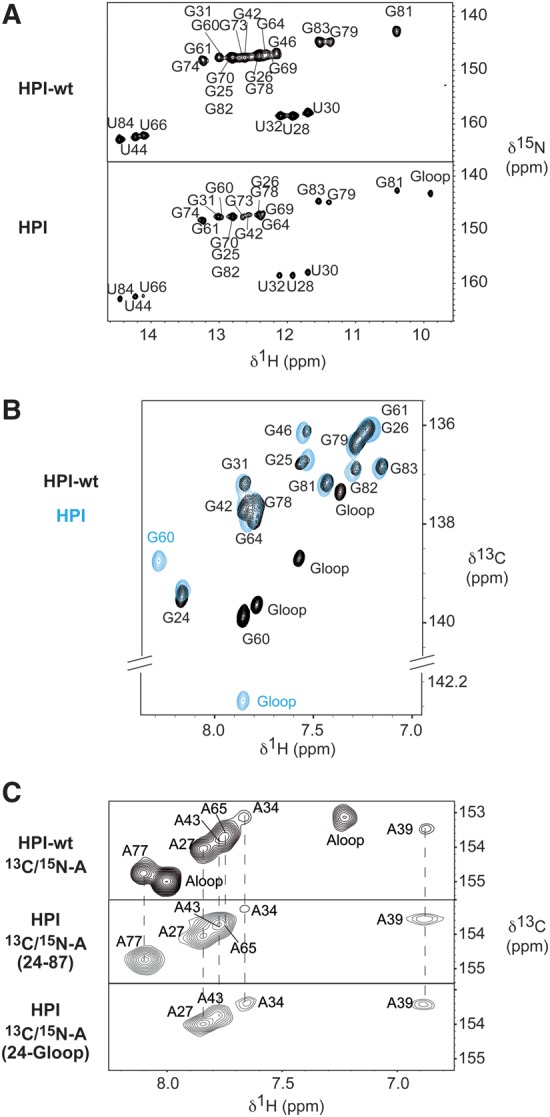
HPI-wt and HPI adopt the same conformation. (A) 1H–15N HSQC spectra showing imino protons region of HPI-wt (top) and HPI (bottom). All imino protons were assigned via sequential nuclear Overhauser effects (NOEs) observed in 2D-NOESY experiments. In the HPI construct, imino protons of the UUCG apical loop resonate at the expected chemical shifts typically observed, which ensures a proper folding of the RNA (Varani et al. 1991; Molinaro and Tinoco 1995). (B) 1H–13C HSQC spectra showing the aromatic H8-C8 of selectively labeled [13C/15N-G] HPI-wt (black) and HPI (blue), recorded at 30°C. (C) 1H–13C HSQC spectra showing the aromatic H2–C2 correlations of the selectively labeled [13C/15N-A] HPI-wt (top), the selectively labeled [13C/15N-A] HPI (middle), and the segmentally and selectively labeled [13C/15N-A(G24-G60)] HPI (bottom). HPI was selectively labeled at all A positions (middle), and segmentally and selectively labeled at A positions in the G24-G60 segment (bottom). Assignment of aromatic H2–C2 of HPI-wt had been previously reported (Lebars et al. 2010). Assignment of HPI was based on analysis of NOESY spectra recorded at 4°C, 10°C, 15°C, 20°C, 30°C in 90/10 H2O/D2O and at 20°C and 30°C in 99.9% D2O. Spectra were recorded in 50 mM sodium phosphate buffer at pH 6.2.