
FIGURE 5.
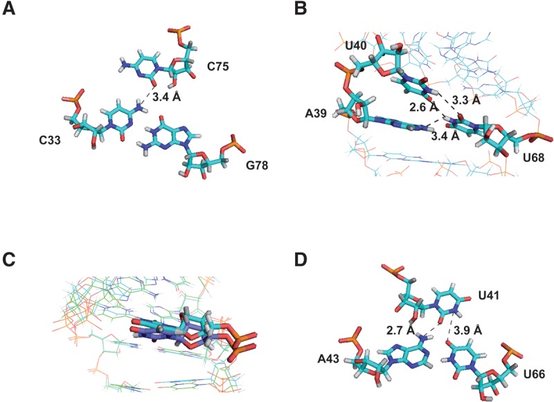
Hydrogen bond networks. (A) Base triple involving C33:G78:C75 in the free-Mg2+ structure. (B) Zoom on A39, U40, and U68 residues in the free-Mg2+ structure. (C) Comparison of the U41 residue from Mg2+-free (dark blue) and Mg2+-bound structures (light blue). (D) Base triple involving A43:U66:U41 in the 3 mM Mg2+ model. Distances consistent with the formation of hydrogen bonds are displayed. Broken lines indicate possible hydrogen bonds.