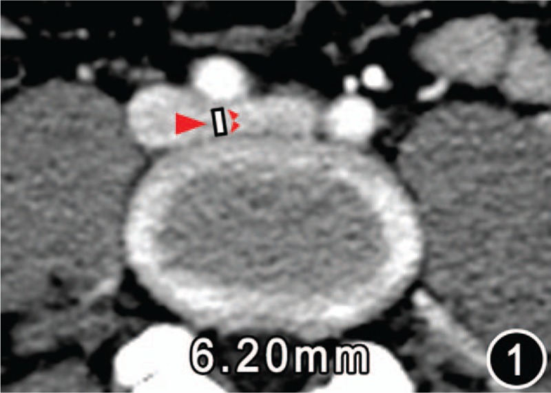
Figure 1.

CT image of a female of age 40 years of control group. The map displayed the diameter (white line) of the left iliac vein tunnel (IVT, big red arrow) 6.20 mm, which was measured on the central cross-sectional CT image of the left iliac vein stretching across the anterospine from right to left. The anterior border of the IVT is the posterior wall of the right iliac artery (the front small red arrow); the posterior border is the anterior margin of the vertebral body (the rear small red arrow).
