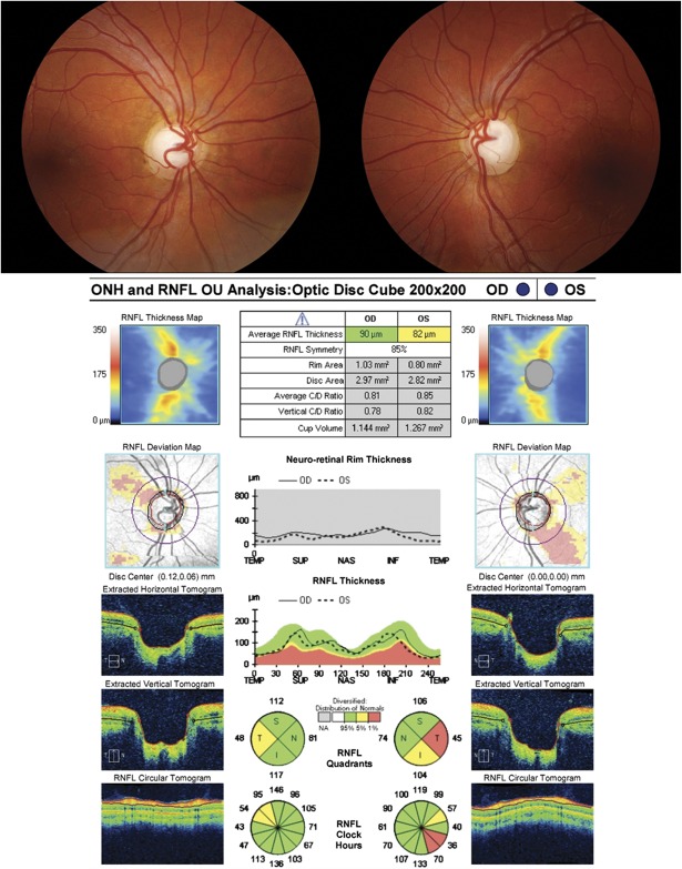
FIG. 12.
A large optic nerve can give the appearance of an enlarged cup-to-disc ratio. This patient was referred for evaluation of presumed glaucomatous optic neuropathy. Fundus photography demonstrates large optic nerves with large cup-disc ratios. OCT of the optic nerve demonstrates a large disc area of almost 3 mm2 with a relatively normal average RNFL thickness. Note that the abnormal RNFL probability map for the superior arcuate sector in the right eye and the inferior arcuate sector in the left eye are due to the more vertical angle of entry of these arcuate bundles, which is evident on the color thickness map plot and the vertical angle of entrance of the arterial branches corresponding to these bundles. This confirms the diagnosis of megalopapilla, which causes an enlarged cup-to-disc ratio in the absence of true glaucomatous optic neuropathy. OCT, optical coherence tomography; RNFL, retinal nerve fiber layer.

