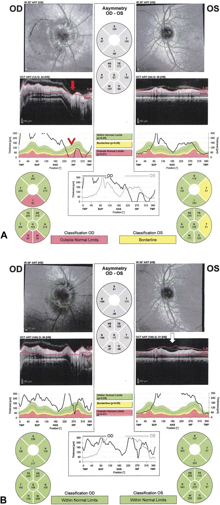
FIG. 4.

Spectralis OCT with artifacts in the RNFL thickness. A. this patient presented with bilateral asymmetric papilledema with Frisen-grade 3 disc edema of the right optic nerve and very mild disc edema of the left optic nerve. Segmentation error of the RNFL is seen on the B-scan of the right eye (red arrow), which causes a nonphysiologic decrease in the RNFL thickness toward 0 μm inferiorly on the RNFL temporal-superior-nasal-inferior-temporal thickness plot (red arrowhead). B. A repeat OCT 1 month later shows less segmentation artifact of the RNFL in the right eye. However, the left eye at 1 month shows a significant thickening RNFL thickness due to errors in identifying the internal limiting membrane due to prominent vitreoretinal interface opacity, which can be seen on the B-scan (white arrow). OCT, optical coherence tomography; RNFL, retinal nerve fiber layer.
