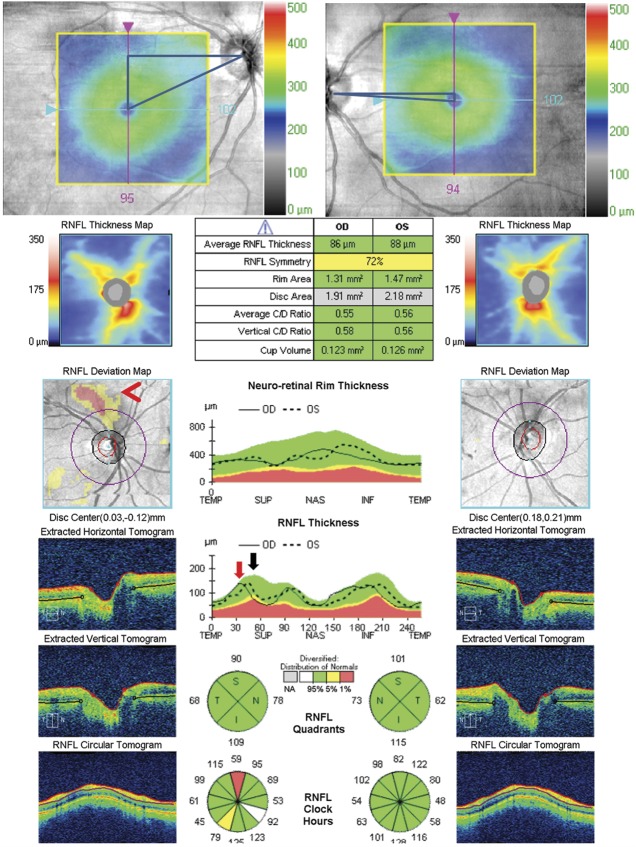
FIG. 7.
Cyclotorsion of the eyes will introduce a shift of the RNFL peaks with respect to the normative database RNFL TSNIT profile that can lead to false-positive probability plots of both TSNIT and RNFL when compared to the normative database. This patient had a right fourth nerve palsy causing significant excyclotorsion of the right eye, which was measured at 17° on double
Maddox rod. The excyclotorsion of the right eye can be seen on the OCT macular thickness map superimposed on the OCT fundus image and can be quantified by triangulation (top images). This introduced an artifactual superior arcuate area of thinning on the probability map with respect to the normative database (red arrowhead) because of the temporal shift of the superotemporal peak, which can also be seen on the TSNIT plot (red arrow indicates shifted peak, black arrow indicates the normal location of the superotemporal peak). In addition, the magnitude of the shift of the peak compared to the normative database equaled the amount of excyclotorsion measured by triangulation on the fundus photo and clinically on double Maddox rod. OCT, optical coherence tomography; RNFL, retinal nerve fiber layer; TSNIT, temporal-superior-nasal-inferior-temporal.

