
FIG. 9.
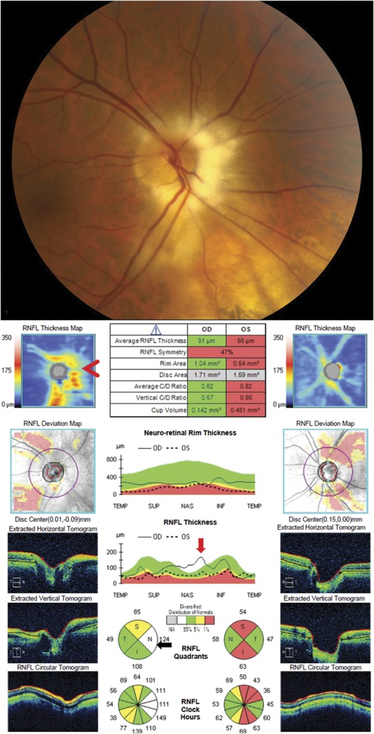
Artificially increased RNFL thickness from myelinated nerve fiber layer. This patient has primary open-angle glaucoma resulting in optic nerve cupping and thinning of the RNFL. The prominent nasal myelinated nerve fiber layer in the right eye results in a thickened RNFL that obscures thinning of the RNFL from the patient's underlying glaucoma. The RNFL thickness map shows the normally positioned superotemporal and inferotemporal arcuate bundles and also an irregular elevated RNFL nasal to the disc (red arrowhead) due to the myelinated nerve fiber layer. The myelinated nerve fiber layer leads to an elevated nasal peak on the temporal-superior-nasal-inferior-temporal plot (red arrow), which causes the nasal RNFL thickness to be thicker than the general population and is therefore shown in white on the RNFL quadrant analysis (black arrow). RNFL, retinal nerve fiber layer.