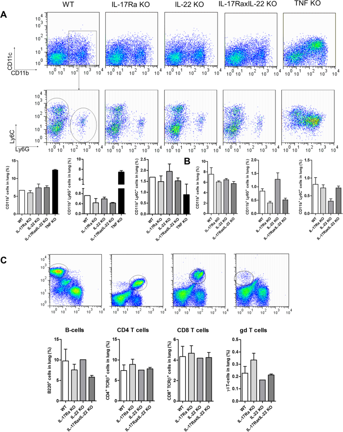
Figure 6. M. tuberculosis induced lung inflammatory cell infiltration in the absence of IL-17RA and IL-22.
Mice deficient for IL-17RA, IL-22, both IL-17RA and IL-22, and TNFα mice as well as wild-type mice were infected with M. tuberculosis (H37Rv, 1000 CFU/mouse i.n.). Inflammatory lung infiltrating cells were analysed by flow cytometry, and bar graphs show data from 2 to 3 individual mice per group, and 2 pools of 2 TNFα-deficient mice, at 1 (A) or 2 months (B,C) post-infection, expressed as mean +/− SEM. Representative dot plots of CD11b+ and CD11c+ cell populations, and Ly6G+ and Ly6C+ cells gated on CD11b+ cells are shown (A). In (C), the gating strategy and relative populations of B220+, CD4+, CD8+ and γδ T lymphocyte populations are shown.

