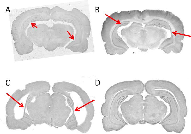
Figure 3.
Histological (Nissl-stained) examples of NVHL (A-C) and sham (D) brains from adult animals. A, NVHL brain with minimal cell loss and disorganization, indicated by arrows. Anterior-posterior (AP) level is approximately −4.5 mm relative to bregma (Paxinos and Watson, 1988). B, NVHL brain with moderate cell loss and disorganization, indicated by arrows. Approximate AP level, −5.3 mm. C, NVHL brain with extensive cell loss and disorganization, indicated by arrows. Note the enlarged ventricles and slight lateral cortical thinning. Approximate AP level, −5.6 mm. D, Sham-treated brain showing an intact hippocampus. Approximate AP level, −5.6 mm.

