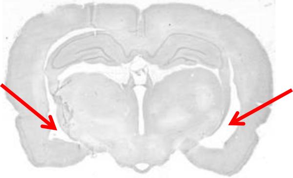
Figure 4.
Nissl-stained example of a sham-treated brain at a more anterior level (approximate AP = −3.8 mm) that may be mistaken for a lesion. This level is just anterior to the juncture of the dorsal and ventral aspects of the hippocampus; at this level, the lateral ventricle is normally enlarged (arrows). This cavity should not be mistaken for a lesion.

