
Figure 3.
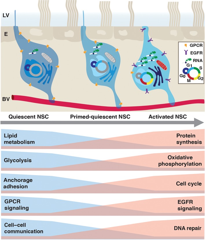
Molecular changes upon stem cell activation. Top: Schema of quiescent (left), primed‐quiescent (middle), and activated (right) adult V‐SVZ NSCs, located between the ependymal cell layer (E) lining the lateral ventricle (LV), and the vascular plexus (BV). Summary of transcriptome data of purified qNSCs and aNSCs at the population and single cell level. EGFR, epidermal growth factor receptor; GPCR, G‐protein coupled receptor; NSC, neural stem cell.