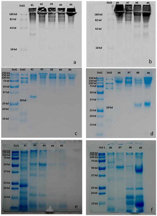
Figure. 6.

SDS-PAGE electrophoresis of the proteins released into the media from Schizosaccharomyces yeasts. Glycoprotein visualization was performed using a two step procedure. The glycoproteins were first stained on electrophoretic gel with Pro-Q Emerald 488 glycoprotein gel stain kit (6 a-b). Then, the proteins were stained in the same gel with Bio safe Coomassie (6 c-d). The pool of proteins released in the media have been loaded in the gel after enzymatic de-glycosylation with PNGase F and then stained in the gel with Bio safe Coomassie (6 e-f). Std1: Blue precision plus molecular weight standard; Std2: CandyCane molecular weight standard.
