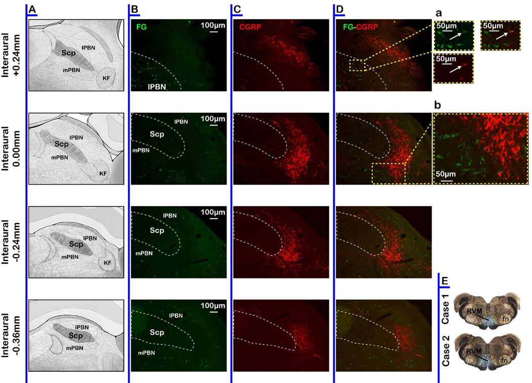
Fig. 10. CGRP-ir neurons in PB do not project to the RVM.
A Schematic representation of the PB. Immunohistochemical label for FG (B, green), CGRP (C, red) and overlap (D) in the PB. Da. Inset showing higher magnification view of the only CGRP/FG double labeled neuron found in PB. Db. Inset showing higher magnification view illustrates segregation of CGRP-ir neurons from RVM-projecting neurons.

