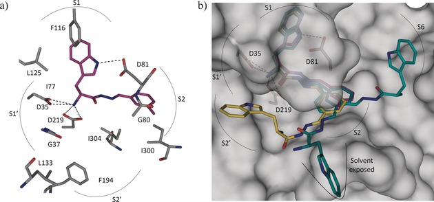
Figure 5.

a) X‐ray crystal structure of endothiapepsin co‐crystallized with bis‐acylhydrazone 13 (PDB ID: 5HCT). b) Superimposition of the crystal structure (violet) and modeled structures (yellow and cyan) of 13.33

a) X‐ray crystal structure of endothiapepsin co‐crystallized with bis‐acylhydrazone 13 (PDB ID: 5HCT). b) Superimposition of the crystal structure (violet) and modeled structures (yellow and cyan) of 13.33