
Figure 1.
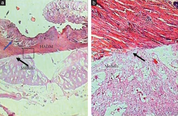
Surgical pathology analysis of lesion in a fetus which underwent prenatal correction of a meningomyelocele‐like defect using human acellular dermal matrix (HADM). (a) HADM adhered to skin (blue arrow) and to neural tissue (black arrow) (hematoxilin‐eosin stain; original magnification, × 16). (b) Magnification of the square section outlined in part (a), showing ingrowth of cells from the medulla (black arrow) into the HADM (hematoxilin‐eosin stain; original magnification, × 100). Reproduced with permission from Sanchez e Oliveira et al. 29.