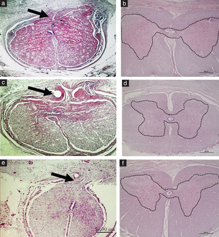
Figure 2.

Surgical pathology specimens of fetal sheep medulla after repair of a spinal defect using two different techniques. (a,c,e) Standard neurosurgical multilayer repair: arrows show adherence of medullar tissue to the scar (meningoneural adhesion). (b,d,f) Skin‐over‐biocellulose technique using biosynthetic cellulose. Images show preservation of the medullar architecture; dashed lines outline the gray matter. Note absence of this line in the neurosurgical group, indicating disruption of the medullary tissue. Reproduced with permission from Herrera et al. 35.
