
Figure 1.
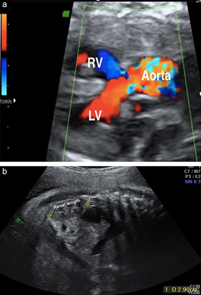
Prenatal ultrasound image in a 35‐week fetus, showing an overriding aorta (a) and a small kidney (length, 29.0 mm vs reference length at 35 weeks of 33.1 mm (3rd percentile) to 51.2 mm (97th percentile)) with poor corticomedullary differentiation (b). LV, left ventricle; RV, right ventricle.