
Fig. 4.
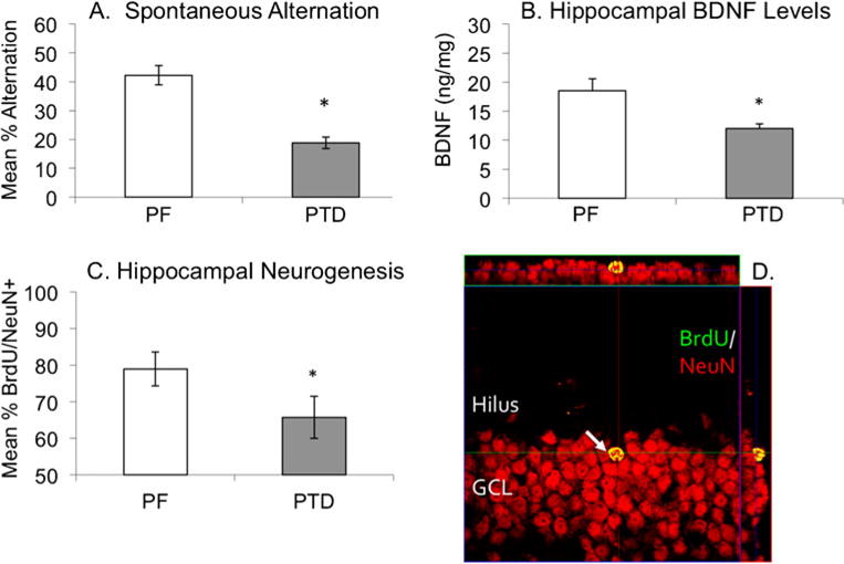
Confocal microscopy image (with orthogonal views) of Bromodeoxyuridine+cells (BrdU+; green) in a PTD brain co-localized with the cells immune positive for the neuronal marker NeuN (Neuronal Nuclei; red) in the subgranular zone (SGZ) of the dentate gyrus. PTD treatment produces a significant deficit in spontaneous alternation (a), chronically decreases hippocampal brain-derived neurotrophic factor (BNDF; b) and reduces hippocampal neurogenesis (c). Co-localized labels of BrdU/NeuN appear yellow and can be confirmed in all three projections (d)