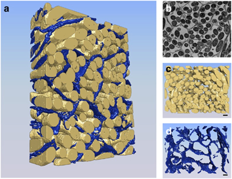
Figure 2.
Three-dimensional reconstruction of M. xanthus microchannel structure through FIB/SEM analysis. M. xanthus swarms were fixed in glutaraldehyde, resin-embedded and stained with Ruthenium Red for microchannel visualization. Blocks were analyzed by SEM that scans the block surface, then the focused ion beam (FIB) oblates 20 nm of material before scanning again. In total, 250 scans were obtained, spanning 5 μm in depth. (a) The entire 3D reconstruction, with cells shown in yellow and channels in blue pseudocolor (Supplementary Movie S3). (b) One of 250 slices captured during the FIB/SEM data collection shows an arrangement of cells within microchannels similar to observations with TEM. Panel (c) shows the 3D reconstruction of cells only and panel (d) shows the 3D reconstruction of microchannels only. Scale bar, 1 μm.

