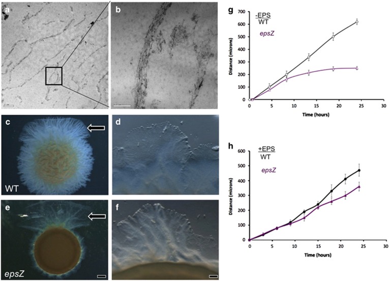
Figure 4.
Purification of EPS microchannels and rescue of swarm migration. (a, b) EM analysis of sodium dodecyl sulfate-purified EPS shows that the purified EPS has a filamentous structure similar to the native conformation of cells within microchannels. (c–f) M. xanthus cells were incubated on a 0.5% agar surface in 5 μl aliquots of cells (~108 cells) adjacent to EPS extracts (arrow, top). (c, d) Wild-type migration occurs in all directions, but is promoted in the direction of the added EPS. (e, f) epsZ- mutant migrates only in the direction of the added EPS. (d) Magnified view of wild-type migration into the EPS-coated surface (see Supplementary Movie S4), (f) epsZ- migration into the EPS-coated surface (see Supplementary Movie S5). (g) Quantification of swarm migration in the absence of EPS extracts shows a steady progression of wild-type swarms (black line), whereas epsZ- swarm migration (purple line) is non-processive. In the presence of EPS extracts (h), however, swarm migration of epsZ- is rescuable. Scale bars represent 1 μm, 0.5 μm, 1 mm and 100 μm.

