Figure 8.
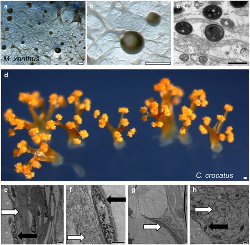
Presence of EPS channels in the 3D structures of Myxobacteria. Comparison of fruiting bodies in (a–c) M. xanthus and (d–h) C. crocatus. (a, b) Stereomicroscopy of M. xanthus grown under conditions that promote both branch migration and fruiting body formation, in which branches connect to spherical fruiting bodies. (c) TEM microscopy of M. xanthus cells within fruiting bodies, showing spores surrounded by layers of EPS. (d) Stereomicroscopy of a field of C. crocatus fruiting bodies, observable as intricately branched orange structures about ~1 mm in height and diameter. The thickness and number of branches varies widely, but the size of each spore sporangiole is similar. (e) TEM analysis of the base of the stalk structures reveals the presence of thick microchannels (white arrows) and several electron-dense cells (black arrows). (f) Further up the stalk, a single cell within a microchannel. (g) A branch tip, where the sporangiole has broken free of the supporting branch. (h) Within the sporangiole, a honeycomb-like organization of filaments surround spores. Scale bars: white, 0.1 mm and black, 1 μm.

