Figure 8.
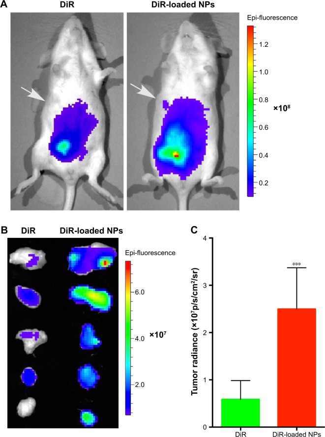
(A) Representative NIRF images of mice (24 h p.i.) injected with DiR solution or DiR-loaded NPs. The white arrows indicate the tumors. (B) Ex vivo NIRF image of tumors after treatment with DiR solution or DiR-loaded NPs (n=5). (C) Quantification of tumor fluorescence showed in panel B (***P<0.005, n=5).
Abbreviations: NIRF, near-infrared fluorescence; NP, nanoparticle; p.i., post-injection.

