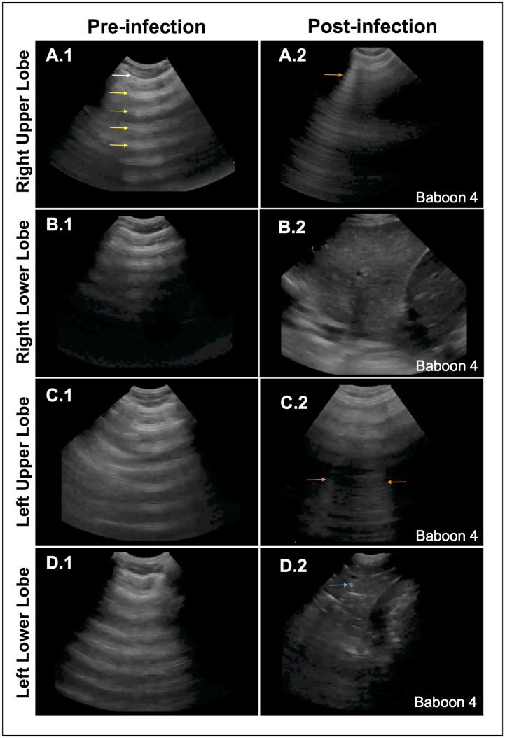
Fig 5. Ultrasonography diagnosis of pneumonia.
Representative images of baseline ultrasonography (A.1-D.1) demonstrate normal lung findings: normal pleura (white arrow) with reverberations of the pleura, or A-lines, seen in the air-filled lung (yellow arrows). The end-of-experiment images demonstrate different pathologic findings. The right upper lobe (A2) shows a discrete, vertical hyperechoic line, or B-line (orange arrow), a type of reverberation artifact due to interlobular septal edema. The right lower lobe (B2) shows a densely consolidated lobe that has similar echogenicity as the liver (“hepatization”) due to replacement of lung air with fluid. The left upper lobe (C2) shows a few B-lines (orange arrows) due to interstitial edema. The left lower lobe (D2) shows a densely consolidated lobe with distinct white speckled areas that are air-bronchograms due to the air-water interface in the terminal bronchioles (blue arrow). A small pleural effusion (*) is also seen.

