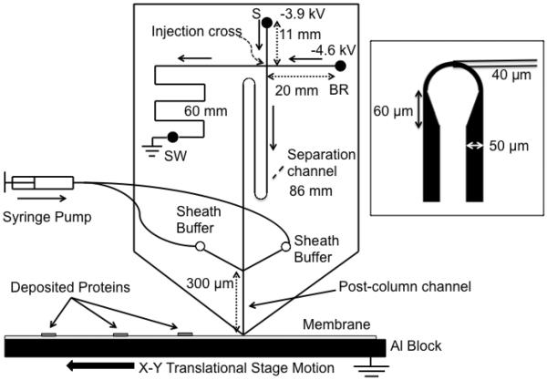
Figure 1.
Microchip overview. Sample was injected using gated injection method. SDS-protein complexes separated by size were captured in discrete zones on the PVDF membrane moving beneath the chip outlet to preserve separation information. Sieving media was pumped through the sheath channels to ensure a stable current. Channel lengths are indicated by double arrow lines and direction of flow during separation is indicated by solid, single arrows. Separation channel was between the injection cross and the end of the channel, and length was 86 mm. Drawing is not to scale. 300 μm post channel was drawn long for clarity. The inset figure shows the asymmetric turn used to reduce geometric dispersion.

