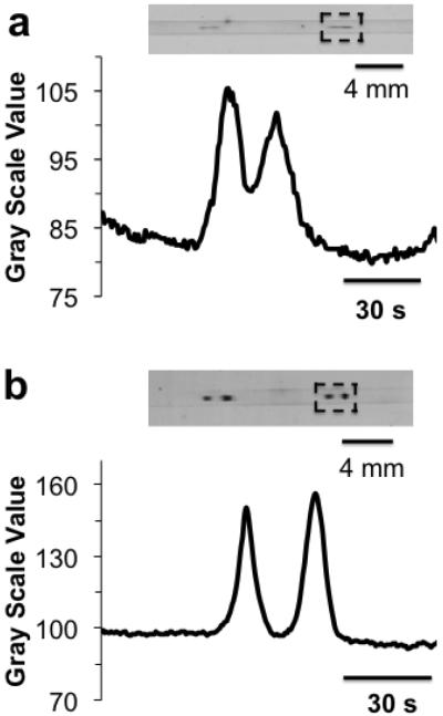
Figure 3.
Western blot assay of ERK1/2 from Jurkat cell lysate with different sample preparation. (a) 2.4 mg/mL cell lysate (results from duplicate injections are shown in blot image) and (b) diluted 1:10 (v:v) with 20 mM phosphate buffer pH 7.5 so that final concentration is 0.24 mg/mL. Traces below each membrane were line scans through membrane with x-axis converted to separation time. Region of interest is boxed with black dashed lines. Sample was injected for 10 s at 390 V/cm using gated injection. Separation field was 400 V/cm over 8.6 cm separation distance.

