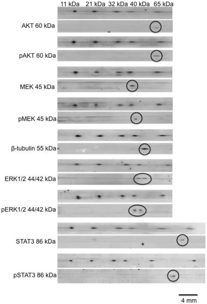
Figure 5.
Multiplexed Western blot assays of Jurkat cell lysate by MCE. 2 μL of 0.2 mg total protein/mL cell lysate was loaded onto the chip. The same sample was injected 9 times with each trace laid on a different membrane. Each membrane was probed with a different antibody as indicated in the figure. For each assay, the membrane was scanned using a fluorescent scanner first at 488 nm/525 nm to detect FITC-protein ladder. After incubating with different antibodies, membranes were scanned at 680 nm/694 nm to detect target proteins. Band for target protein is circled. Expected molecular weights of all the proteins targeted are labeled in the figure.

