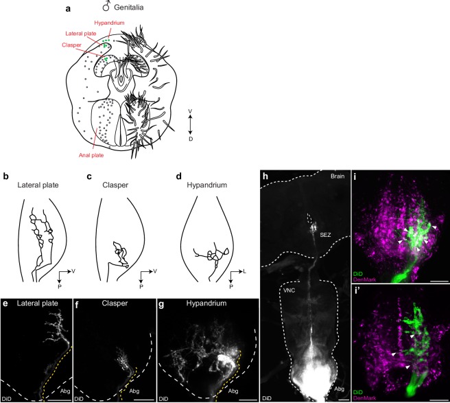
Figure 6. Mechanosensory neurons of the genitalia arborize onto the Abg and brain, and interdigitate with glutamatergic dsx motor neurons in the Abg.
(a) Schematic of male terminalia depicting bristles (right) and bristle topography (dots on left). Dye-filled bristle topography in shown with green dots. (b-d) Schematic of representative lateral plate (b), clasper (c), and hypandrium (d) arborizations in the abdominal ganglion (Abg) of male flies, as previously described (Taylor, 1989). (e-g) Representative images showing topographically distinctive patterns of dye-filled genital neuron arborizations in the male Abg. Maximum intensity z-projections of confocal stacks showing unilateral arborizations of (e) lateral plate, (f) clasper, and (g) hypandrium neurons in the male Abg, which reiterate previously described arborizations (Taylor, 1989). Afferent projections from lateral plate and clasper neurons occupy the same dorso-ventral area but differ in their anterior to posterior positions within the Abg, with clasper neurons ending more posteriorly than lateral plate neurons (b,c and e,f). See also Videos 6,7. Hypandrium neurons typically exhibit a unique contralateral arborization pattern within the Abg (d,g). See also Video 8. (h) A subset of clasper neurons project to the brain. Unilateral dye-fill of clasper neurons together with extended incubation (10 days) reveals single afferent axon (per hemisphere) that transverses the VNC and terminates in the subesophageal zone (SEZ) of the brain. DiD dye-filled arborizations shown in white. (e-h) DiD dye-filled arborizations shown in white. Boundaries of Abg and brain shown with dotted white line. Afferent projections of dye-filled neurons traced with dotted yellow line. D, dorsal, L, lateral, P, posterior, V, ventral. Scale bar = 25 μm. (i) Arborisations of clasper, lateral plates and hypandrium neurons interdigitate with dsx/vGlut dendrites in the adult male Abg. Neurons of all three genital structures were unilaterally dye-filled in males expressing dendritic marker (UAS-DenMark) in dsx/vGlut neurons. Maximum intensity Z-projection of Abg (i) and 10 μm sub-stack (i’) show overt interdigitation (arrowheads) between neurons of all three genital structures and dendrites of dsx/vGlut neurons in the Abg. DenMark shown in magenta; DiD shown in green. Scale bar = 25 μm.

