
Figure 5.
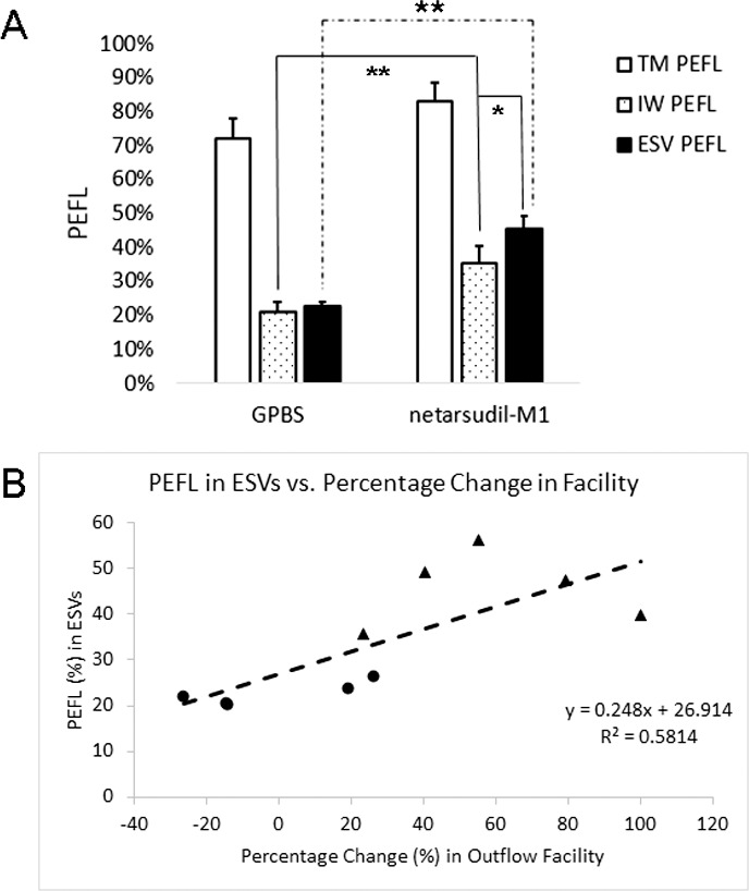
Episcleral vein PEFL and IW PEFL. (A) Netarsudil-M1 treatment induced a significant increase in both ESV and IW PEFL when compared to controls. While ESV PEFL and IW PEFL remained similar to each other in control eyes, ESV PEFL in treated eyes showed a significant increase beyond the increase observed for the IW PEFL (*P < 0.05; **P < 0.01) (n = 5). (B) Episcleral vein PEFL was found to have a positive correlation with percentage change in outflow facility (R2 = 0.58; P = 0.01; solid circles: control eyes; solid triangles: netarsudil-M1–treated eyes).