Figure 1.
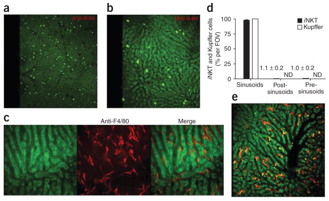
Distribution of iNKT cells and Kupffer cells in the hepatic microvasculature. Spinning-disk confocal intravital microscopy of the vasculature of Cxcr6gfp/+ and BALB/c mouse livers. (a,b) CXCR6+ cells in the liver of Cxcr6gfp/+ mice. (c) Liver-specific Kupffer cells (red) labeled with phycoerythrin-conjugated anti-F4/80 in the hepatic sinusoids of a BALB/c mouse. (d) GFP+ cells and F4/80+ cells in the sinusoids, post-sinusoidal venules and pre-sinusoidal venules of Cxcr6gfp/+ mouse livers (n = 7 mice) in 21 FOV (iNKT cells) or 10 FOV (Kupffer cells). Numbers in graph indicate percent iNKT cells per FOV for bars not visible; ND, not detected. (e) Dextran conjugated to tetramethylrhodamine (2 megadalton; red) and polychromatic microspheres (green and yellow) bound to Kupffer cells. Original magnification, ×4 (a and iNKT cells in d), ×10 (b,e and Kupffer cells in d) or ×20 (c). Data are representative of more than three independent experiments (a–c,e) or seven experiments (d; error bars, s.e.m.).

