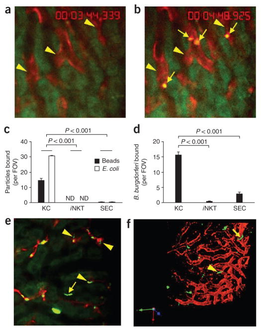
Figure 2.
Binding capacity of Kupffer cells, iNKT cells and SECs for beads and bacteria. (a,b) Microscopy of red Kupffer cells (arrowheads) before (a) and after (b) intravenous injection of polychromatic microspheres (arrows, b) into BALB/c mice. (c,d) Quantification of the binding of beads or E. coli (c) or B. burgdorferi (d) to Kupffer cells (KC), iNKT cells or SECs in Cxcr6gfp/+ mice given intravenous injection of polychromatic microspheres, or E. coli (c) or B. burgdorferi (d) expressing GFP or labeled with the red fluorescent nucleic acid stain Syto 60. (e) Binding of B. burgdorferi to the endothelium (arrow) as well as to Kupffer cells (arrowheads) in the hepatic sinusoids of a mouse treated as described in d (differences in activity, Supplementary Video 5). (f) Microcopy of B. burgdorferi in the liver of a mouse treated as described in d; yellow arrowhead indicates a GFP+ spirochete in the process of emigrating, and yellow arrow indicates a spirochete that has left the vasculature. Original magnification, ×20 (a,b,e,f). P values (c,d), Bonferroni’s multiple-comparison test. Data are representative of more than three independent experiments (a–e) or two experiments (f).

