Figure 3.
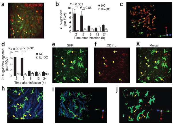
Ingestion of B. burgdorferi by Kupffer cells and Ito cells. (a) Visualization of the hepatic vasculature of a Cxcr6gfp/+ mouse at 2 h after the administration of GFP-expressing B. burgdorferi (yellow dots, shown interacting with red-labeled Kupffer cells). (b) Association of GFP-expressing or Tomato-expressing B. burgdorferi with Kupffer cells and Ito or dendritic cells (Ito-DC), measured in intravital videos of Cxcr6gfp/+ and Cx3cr1gfp/+ mice, respectively. (c) A z-stack reconstruction of two-dimensional microscopy, showing attachment and ingestion of spirochetes by Kupffer cells (rotation, Supplementary Fig. 1). (d) Total ingested B. burgdorferi confirmed after 360° rotation of z-stack-reconstructed images. (e–g) CD11c−, GFP+ big stellate (Ito) cells (arrows) and relatively small CD11c+GFP+ (dendritic) cells (arrowheads) in Cx3cr1gfp/+ mouse liver. (h) Tomato-expressing B. burgdorferi bound by GFP+ Ito cells in Cx3cr1gfp/+ mouse liver. Arrows indicate associated spirochetes; arrowheads indicate spirochetes out of Ito cells. Vessels were stained with anti-PECAM-1 (blue). (i,j) Ingestion of Tomato-expressing B. burgdorferi by Ito cells, assessed after z-stack reconstruction. Original magnification, ×20 (a,c,e–j). P values (b,d), Bonferroni’s multiple-comparison test. Data are representative of two independent experiments per group with more than four FOV each (a–d,h–j; error bars (b,d), s.e.m.) or two experiments (e–g).

