Figure 7.
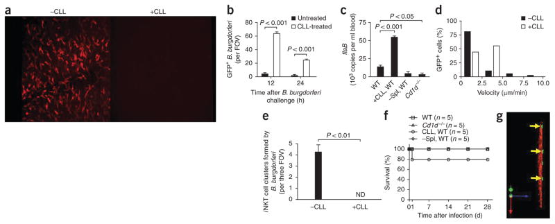
Role of Kupffer cells in B. burgdorferi infection. (a) Kupffer cells labeled with anti-F4/80 before (-CLL) and 24 h after (+CLL) intravenous injection of CLLs (administered 24 h before B. burgdorferi infection to deplete mice of Kupffer cells). Original magnification, ×10. (b) Spirochetes remaining in the liver 12 h and 24 h after B. burgdorferi injection in untreated mice (-CLL) and CLL-treated mice (+CLL). (c) Spirochetes remaining in the blood 3 d after B. burgdorferi injection in untreated, CLL-treated and splenectomized (–Spl) wild-type mice and in Cd1d−/− mice, assessed by quantitative PCR analysis of B. burgdorferi flaB. (d) Distribution of iNKT cell velocities at 5 h after spirochete challenge; nonfluorescent B. burgdorferi were administered to avoid possible confusion of GFP-expressing B. burgdorferi with GFP+ iNKT cells in CLL-treated mice. (e,f) Formation of iNKT cell clusters at 24 h after B. burgdorferi infection. (e) Mortality of untreated, CLL-treated and splenectomized (-Spl) wild-type mice and of Cd1d−/− mice after B. burgdorferi infection. (g) Migration of GFP-expressing spirochetes (arrows) out of microvasculature (space of Dissé) in a CLL-treated mouse. Original magnification, ×20. P values Bonferroni’s multiple-comparison test (b) or Student’s t-test (c,e). Data are representative of three experiments (a), more than two independent experiments per group (b,d,e,g), one experiment with duplicate pooled results of three mice (c) or one experiment with five mice per group (f; error bars (b,c,e), s.e.m.).

