Figure 3.

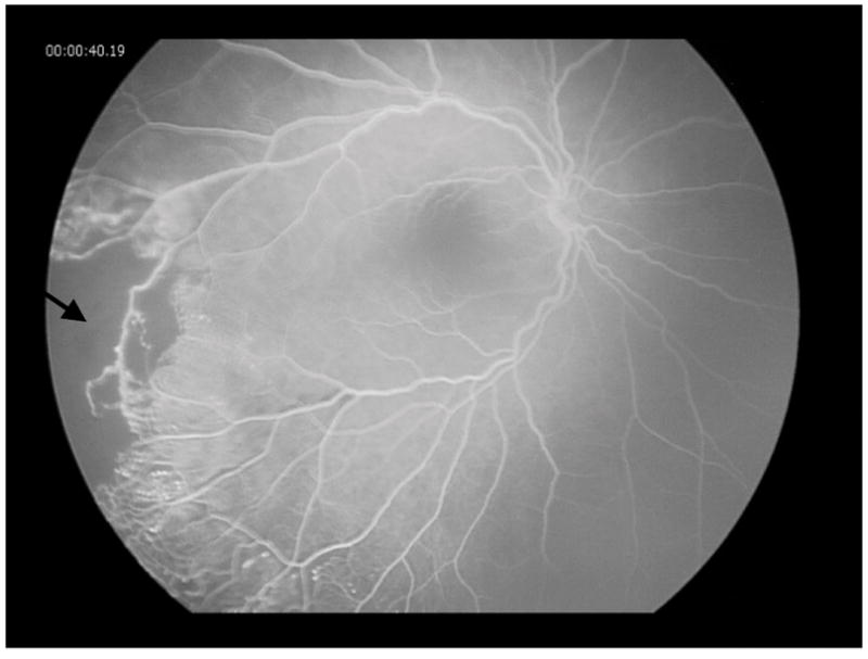
Fluorescein angiography of the patient’s left eye showing vascular telangiectasia, microaneurysms, collateral vessels, and severe retinal non-perfusion (arrow) with fluorescein leakage caused by neovascularization of the disc and retina.

Fluorescein angiography of the patient’s left eye showing vascular telangiectasia, microaneurysms, collateral vessels, and severe retinal non-perfusion (arrow) with fluorescein leakage caused by neovascularization of the disc and retina.