. 2016 Nov 4;8(4):475–480. doi: 10.4055/cios.2016.8.4.475

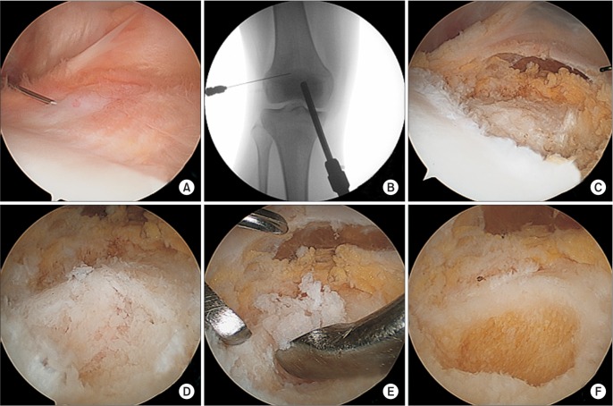
Fig. 4. (A) Arthroscopic image demonstrating a slightly elevated bony lesion at the bottom of the suprapatellar recess near the superior articular margin of the lateral femoral condyle with surrounding minimal hypertrophic synovium. (B) C-arm image taken with a 20-gauge needle held in place to confirm the correct position of the lesion. (C) Removal of the synovium over the lesion using a punching forceps and an electrocautery device. (D) Grossly reddish dense nidus of the osteoid with interconnected trabeculae observed after removal of the sclerotic rim. (E) Excision of the lesion using a small osteotome and curettes. (F) Arthroscopic confirmation of the complete removal of the reactive bone walls performed using a motorized burr.

Fig. 4