Figure 7.

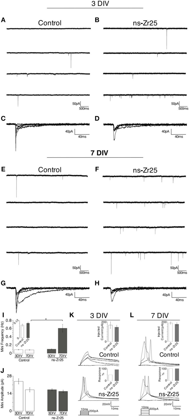
Electrophysiological recordings from cultured hippocampal neurons on flat glass (Control) or the nanostructured zirconia surface (ns-Zr25). Hippocampal neurons were plated on glass (Control) or nanostructured zirconia surfaces (ns-Zr25). Electrophysiological recordings (see methods for details) were done after 3 (A–D,K) or 7 (E–H,L) days of in vitro maturation on these surfaces. Exemplary miniature current traces recorded from neurons plated on Control or ns-Zr25 surfaces after 3 DIV are shown in panels (A) and (B), and after 7 DIV in panels (E) and (F). Bars in graph (I) represent the corresponding mean frequency of miniature postsynaptic currents (mPSCs). At 3 DIV no significant difference between Control (white bars) and ns-Zr25 (gray bars) was found, even if a trend (see also the inset) of an increased frequency in the ns-Zr25 condition starts to emerge (Control = 0.087 ± 0.008, n = 8 cells; ns-Zr25 = 0.101 ± 0.008, n = 14 cells; p > 0.05; Wilcoxon rank-sum test, error bars are sem). This tendency stands out at 7 DIV, at this stage a significant increase in mPSCs frequency in neurons grown on ns-Zr25 surfaces was found (Control = 0.082 ± 0.009, n = 8 cells; ns-Zr25 = 0.606 ± 0.094, n = 10 cells; p < 0.05; Wilcoxon rank-sum test, error bars are SEM). Representative events from exemplary neurons are overlapped in panels (C,D,G,H) to show the variability in shape and amplitude in the different conditions. (J) The bar panel displays the mean of the amplitude mPSCs in the different conditions. The obtained data show a trend in the Control condition; with a higher mean amplitude of the miniatures in immature neurons (3 DIV) and a decrease over maturation (7 DIV) as expected from previous reports (Bose et al., 2000). On ns-Zr25 stable mean amplitude was observed over maturation with a value in the range of the mean value seen for the mature neurons grown in the Control condition at 7 DIV (3 DIV mean amplitude: Control = 23.76 ± 2.09; ns-Zr25 = 17.11 ± 0.8; 7 DIV mean amplitude: Control = 17.04 ± 1.68; ns-Zr25 = 15.8 ± 0.1). Panels (K) and (L) represent exemplary membrane voltage recordings from individual neurons cultured in different conditions. The insets display the injected current thresholds and the percentage of responding cells. When triggered to fire action potentials, by current injections steps (injected current protocol scheme on the bottom), (K) young neurons (3 DIV) cultured on ns-Zr25 surfaces demonstrate an enhanced excitability compared to neurons maintained in Control condition. (L) At 7 DIV Control neurons acquired excitability comparable to the ns-Zr25 condition (injected current protocol scheme on the bottom).
