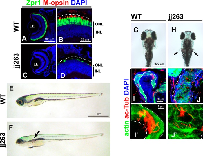
FIGURE 1.
jj263 mutant larvae exhibited photoreceptor degeneration and cystic kidney without curly body axis phenotype. A–D, jj263 mutant larvae showed photoreceptor degeneration in the retina. Retinal sections at 10 dpf of wild-type (A and B) and jj263 mutant (C and D) larvae were immunostained with anti-Zpr1 (an-arrestin, double cone photoreceptors, green) and anti-M-opsin (a cone outer segment marker, red) antibodies. Nuclei were stained with DAPI (blue). In the wild-type retina, cone photoreceptors are localized in the ONL and form the outer segments on apical sides of the ONL. In the jj263 mutant larvae, photoreceptor cells in the ONL were severely degenerated. In contrast, the INL in the jj263 mutant larvae was almost normal. Dotted lines indicate the boundary of retinal pigment epithelia. E–H, lateral (E and F) and dorsal (G and H) views of wild-type (E and G) and jj263 mutant (F and H) larvae at 6 dpf. A cystic kidney phenotype was observed in the jj263 mutant larvae (F and H, arrow). The curly body axis phenotype, a common feature of ciliary mutants, was not observed in the jj263 mutant larvae. I–J′, pronephric cilia are disorganized and shortened in the jj263 mutant larvae. Pronephric cilia in the wild-type (I and I′) and mutant (J and J′) larvae were immunostained with an acetylated α-tubulin antibody (red). Pronephric epithelia in pronephric tubules were stained with phalloidin (green). Nuclei were stained with DAPI (blue). Dotted lines indicate boundaries of pronephric tubules. ONL, outer nuclear layer; INL, inner nuclear layer; LE, lens.

