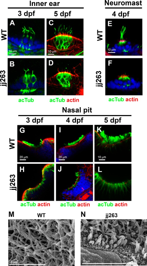
FIGURE 3.

Ciliary defects in sensory organs of the ift122 mutant. A–F, hair cell kinocilium in the inner ear (A–D) and neuromasts (E and F) of wild-type (A, C, and E) and jj263 (B, D, and F) embryos at 3 dpf (A and B), 4 dpf (E and F), and 5 dpf (C and D). Cilia are immunostained with an anti-acetylated α-tubulin antibody (green). Stereocilia of hair cells were stained with phalloidin (actin staining, red). Nuclei were stained with DAPI (blue). Kinocilia of hair cells were disorganized in the ift122 mutant inner ear and neuromast. G–L, cilia in the wild-type (G, I, and K) and ift122 mutant (H, J, and L) nasal pit at 3 dpf (G and H), 4 dpf (I and J), and 5 dpf (K and L). Cilia are immunostained with an anti-acetylated α-tubulin antibody (green). Apical surface of epithelia were labeled with phalloidin (actin staining, red). Nuclei were stained with DAPI (blue). Cilia do not develop in mutant nasal pit at 3 dpf. M and N, ultrastructural analysis of nasal cilia in wild-type and ift122 mutant larvae at 4 dpf. Swollen cilia were observed in the ift122 mutant.
