FIGURE 4.

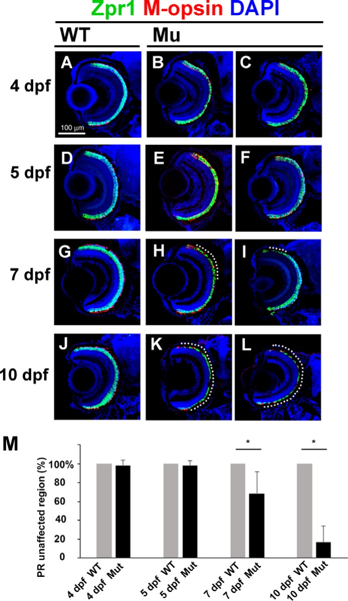
Slow progressive photoreceptor degeneration in the ift122 mutant retina. A–L, ift122 mutant larvae showed slow photoreceptor degeneration in the retina. Retinal sections at 4 dpf (A–C), 5 dpf (D–F), 7 dpf (G–I), and 10 dpf (J–L) of wild-type (A, D, G, and J) and ift122 mutant (B, C, E, F, H, I, K, and L) larvae were stained with anti-Zpr1 (double cone photoreceptors, green), and M-opsin (a cone outer segment marker, red) antibodies. Nuclei were stained with DAPI (blue). No photoreceptor degeneration was observed in the ift122 mutant retina between 4 and 5 dpf. Only small portions of the photoreceptor layer degenerate at 7 dpf (H and I, dotted lines). Most of the photoreceptor layer degenerated by 10 dpf in the ift122 mutant retina (K and L). M, we measured the depth of the degenerated photoreceptor region and calculated the size of the unaffected region. Significant photoreceptor degeneration was observed at 7 and 10 dpf. n = 9–18.
